Eye Library / Eye Anatomy
Eyeball is a spherical structure approximately 2.5 cm in diameter with a pronounced bulge on its forward surface. Even though the eye is small, it serves a very important function that is sense of sight. Vision is arguably the most used of the 5 senses and is one of the primary means that we use to gather information from our surroundings.
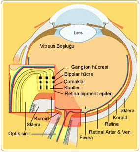
The eye functions like a camera. Each gathers light and then transforms that light into a “picture.” Both also have lenses to focus the incoming light. A camera uses the film to create a picture, whereas the eye uses a specialized layer of cells, called the retina, to produce an image. Later, messages about the focused picture are sent to the brain through the optic nerve and this is how we see.
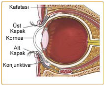
Orbit is the eye socket, which is formed by the cheekbone, the forehead, the temple, and the side of the nose. The eye is cushioned within the orbit by pads of fat.
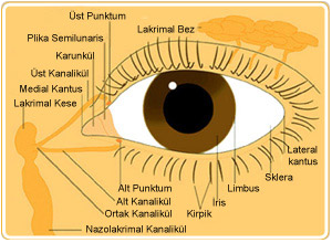
Orbit also contains lacrimal gland that is located underneath the outer portion of the upper eyelid. The lacrimal gland produces tears that help lubricate and moisten the eye, as well as flush away any foreign matter that may enter the eye. The tears drain away from the eye through the nasolacrimal duct, which is located at the inner corner of the eye.
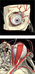
Six extraocular muscles are attached to each eye to help move the eye left and right, up and down, and diagonally.
 The eye is approximately a spherical shell transparent at the front portion and opaque over the remaining eighty per cent of its surface at the back.
At the front of the eyeball, it is continuous with the bulging, transparent cornea. Cornea is a clear layer at the front and center of the eye. In fact, the cornea is so clear that you may not even realize it is there. The cornea is located just in front of the iris, which is the colored part of your eye. The main purpose of the cornea is to help focus light as it enters the eye. If you wear contact lenses, the contact lens rests on your cornea.
The white part of your eye that you see when you look at yourself in the mirror is the front part of the sclera. Sclera is a tough, leather-like tissue, also extends around the eye. Just like an eggshell surrounds an egg and gives an egg its shape, the sclera surrounds the eye and gives the eye its shape. The sclera is also attached to the extraocular muscles, which, in turn, move the eye left and right, up and down, and diagonally.
 The middle layer of the coating of the eye is choroid, a vascular layer lining the posterior three-fifths of the eyeball. The choroid is continuous with the ciliary body and with the iris, which lies at the front of the eye. The innermost layer is the light-sensitive retina.
Several structures, not parts of the eyeball, contribute to the protection of the eye. The most important of these are eyelids, two folds of skin and tissue, upper and lower, that can be closed by means of muscles to form a protective covering over the eyeball against excessive light and mechanical injury. When you blink, the eyelids also help spread tears over the surface of your eye, keeping the eye moist and comfortable
Eyelashes, a fringe of short hairs growing on the edge of either eyelid, act as a screen to keep dust particles and insects out of the eyes when the eyelids are partly closed. The eyelashes help filter out foreign matter, including dust and debris, and prevent it from getting into the eye.
Tears are produced in the lacrimal glands, which are located above the outer segment of each eye. The tears eventually drain into the inner corner of the eye, into the lacrimal sac, then through the nasal duct and into the nose. That is why your nose runs when you cry.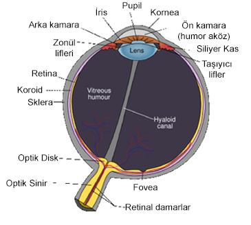 Anterior chamber angle and the trabecular meshwork are located where the cornea meets the iris. The trabecular meshwork is important because it is the site where the aqueous humor drains out of the eye. If the aqueous humor cannot properly drain out of the eye, the pressure can build up inside the eye, causing optic nerve damage and eventually vision loss, a condition known as glaucoma.
Iris is the colored part of the eye. The color of the iris is determined by the color of the connective tissue and pigment cells. Less pigment makes the eyes blue; more pigment makes the eyes brown. The iris is a shutter which automatically controls to some degree the amount of light reaching the retina. Pupil size can change from 2 millimeters to 8 millimeters. This means that by changing the size of the pupil, the eye can change the amount of light that enters it by 30 times. The iris has two muscles to perform its function: The dilator muscle makes the iris smaller and therefore the pupil larger, allowing more light into the eye. The other one is the sphincter muscle makes the iris larger and the pupil smaller, allowing less light into the eye.
Posterior chamber is the fluid-filled space immediately behind the iris but in front of the lens. The fluid that fills this chamber is called the aqueous humor. The aqueous humor helps to nourish the cornea and the lens.
Lens is a clear, flexible structure that is located just behind the iris and the pupil. A ring of muscular tissue, called the ciliary body, surrounds the lens. Together, the lens and the ciliary body help control fine focusing of light as it passes through the eye.
 Vitreous cavity is located behind the lens and in front of the retina. It is filled with a gel-like fluid, called the vitreous humor. The vitreous humor helps maintain the shape of the eye.
Retina has two main parts: the peripheral retina and the macula.
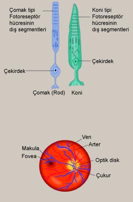
Macula is located in the central part of the retina. It is the area of the retina that is responsible for giving you central vision. The macula is the tiny yellow spot on the retina having 6 mm in diameter where detail focus occurs. There is also more specific part of macula which is named fovea
Fovea contains the greatest concentration of cones. The part of the image that is focused on the fovea is the image most accurately registered by the brain.
 Choroid is the middle layer of the eye between the retina and the sclera. It is mostly made up of blood vessels. The choroid helps nourish the retina. It also contains a pigment that absorbs excess light so preventing blurring of vision.
The retina is primarily made up of 2 distinct types of cells: rods and cones. Rods are more sensitive to light; therefore, they allow people to see in low light situations but do not allow them to see color. There are approximately 125 million rods. They are spread throughout the peripheral retina and function best in dim lighting. Cones allow people to see color but require more light.


