Retinal Tears And Detachment
What is retinal tear and retinal detachment?
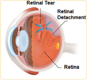
Retinal tear and retinal detachment are part of the same spectrum of disease requiring urgent evaluation and treatment. The retina is a thin layer of nerve cells that lines the inside of the eye. In the young eye, the vitreous is in direct contact with the retina. With aging, the vitreous slowly shrinks and these fine fibers pull on the retinal surface. Usually the fibers break, allowing the vitreous to separate from the retina. In most cases the patient may notice flashes and/or floaters, but vitreous will separate from the retina without complication. Rarely, retinal tears develope. If the retinal tear remains unrecognized and untreated, the fluid can build up under the retina leading to retinal detachment. A retinal detachment may progress quickly and lead to complete loss of vision.
Who is at risk for retinal detachment?
Retinal detachment most often occurs as a result of the natural aging process, but certain people are at higher risk than others. These include high myopic, those with a history of cataract surgery, and those who have recently suffered an ocular trauma. Some types of retinal detachments can run in families.
What are the symptoms of retinal detachment?
The symptoms of vitreous separation, retinal tear, and retinal detachment are similar and sometimes can overlap. On occasion, the patient may notice floaters, flashing lights and loss of peripheral vision which may present as a shadow moving toward the center of one’s field of vision. Floaters and flashes do not always mean that you will have a retinal detachment. But they may be a warning sign and should be evaluated promptly.
How retinal detachment is diagnosed?
Retinal tear and retinal detachment can be recognized during dilated fundoscopic examination, an examination performed by using an ophthalmoscope and magnified condensing lens.
How are retinal tears treated?
If a retinal break can be discovered before a retinal detachment develops it can be treated with the laser to seal the break and prevent a retinal detachment. The laser creates small burns around the edges of the tear, producing scars. These scars seal the borders of the tear and prevent fluids from separating retina from underlying tissues.
How is retinal detachment treated?
Retinal detachment almost always causes blindness unless it is treated. A detached retina is almost always repaired surgically. Surgical options include pneumatic retinopexy, scleral buckling, or vitrectomy. Pneumatic retinopexy involves injecting a gas bubble into the vitreous space in combination with laser surgery or cryotherapy. A scleral buckle is a tiny, flexible band that is placed around the outside of the eyeball to gently push the wall of the eye against the detached retina. Vitrectomy is the surgical extraction of the vitreous humor, reattaching retina and simultaneous replacement with a dissolvable gas bubble or a silicone oil.
Last Updated: October 23, 2024


